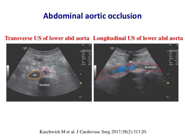
Aorta Branches Ultrasound
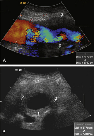
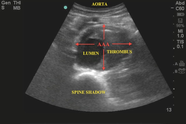
Its the preferred screening method for an abdominal aortic aneurysm a weakened bulging spot in the abdominal aorta the major blood vessel that supplies blood to the body.

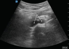
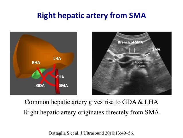
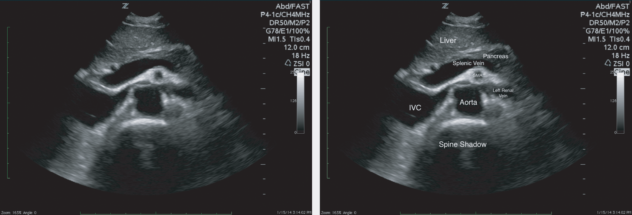
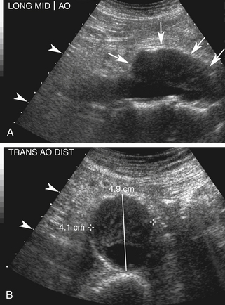
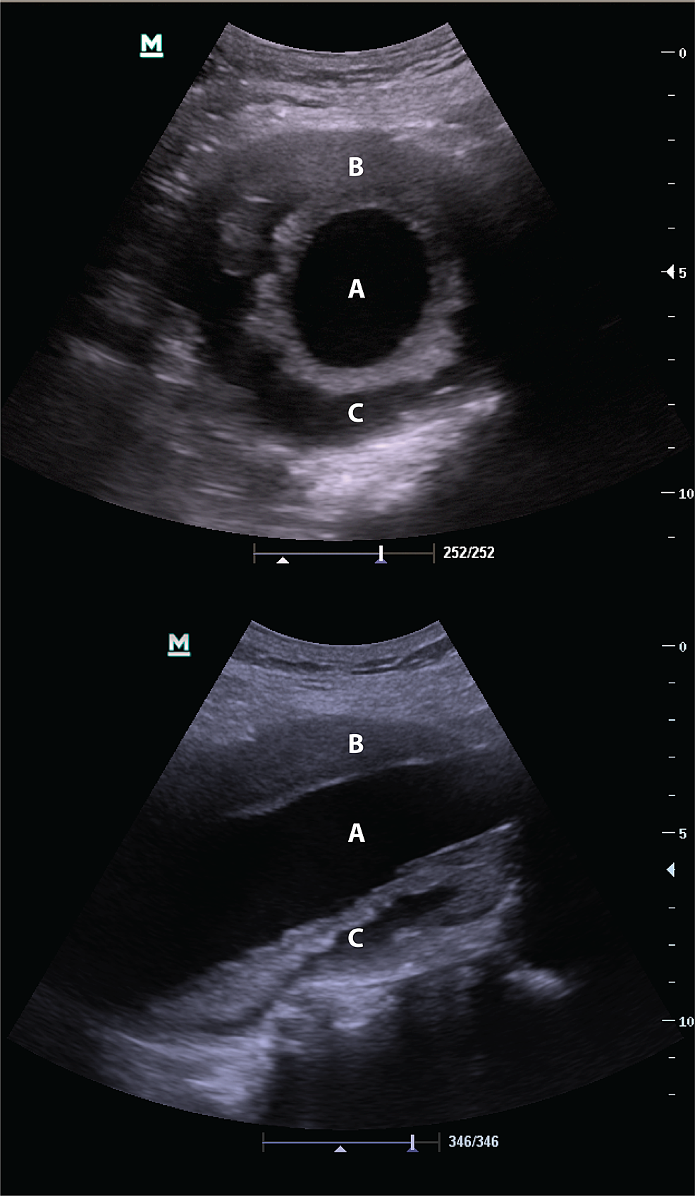
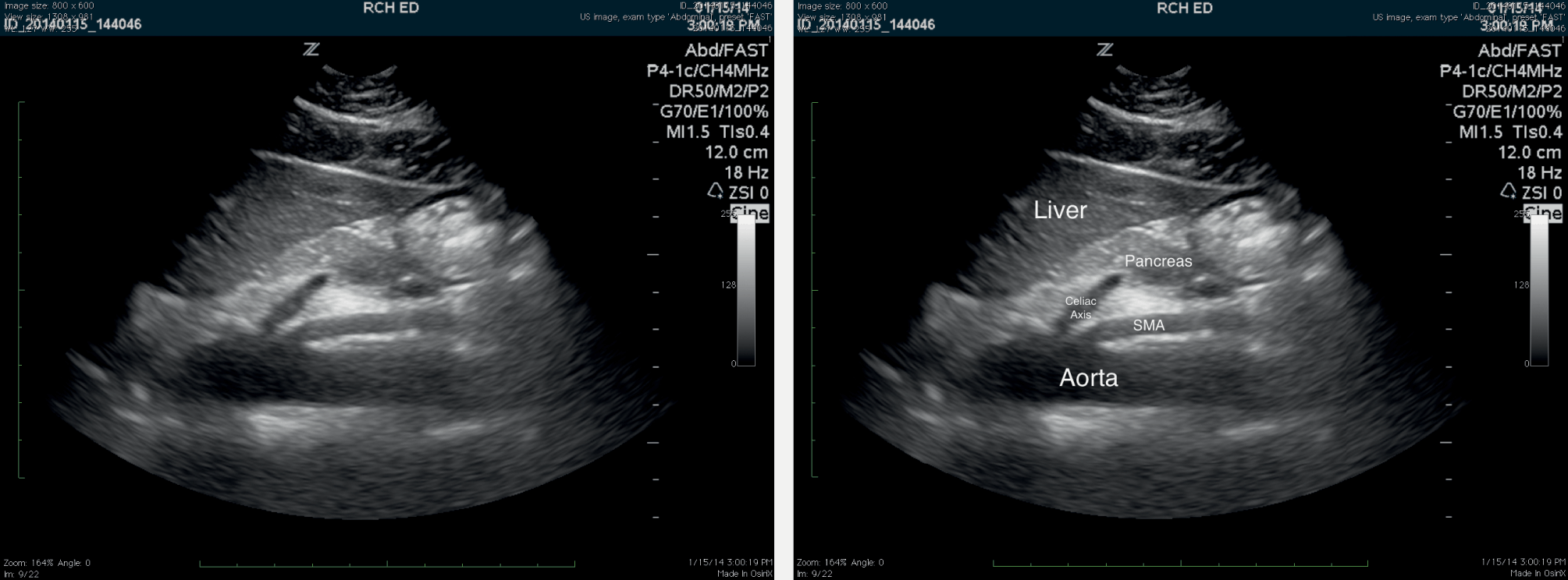
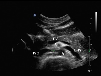
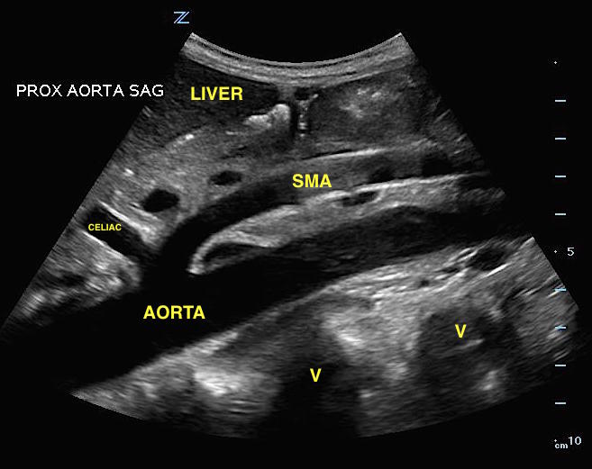
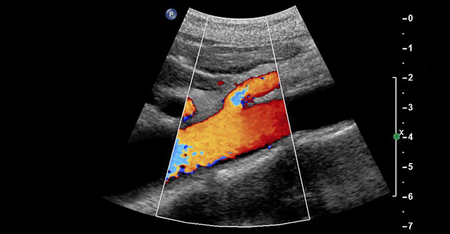
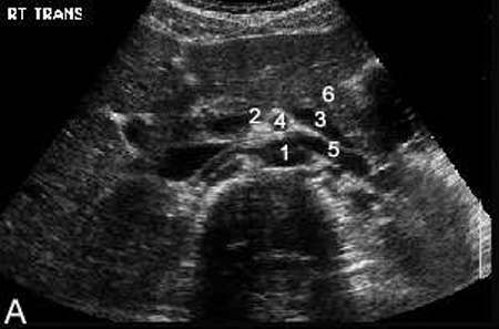
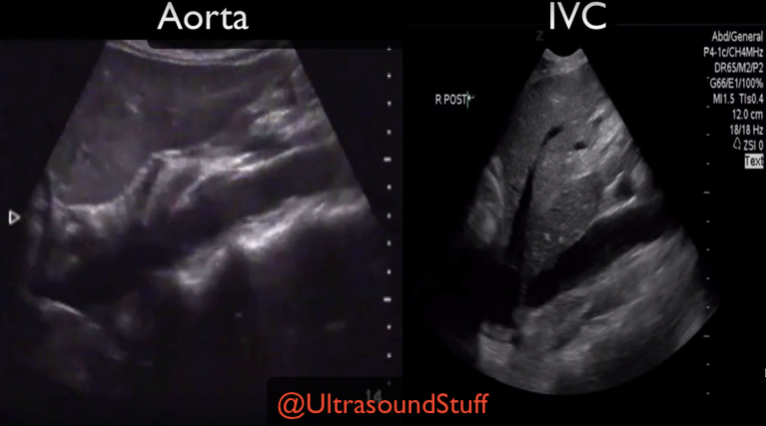
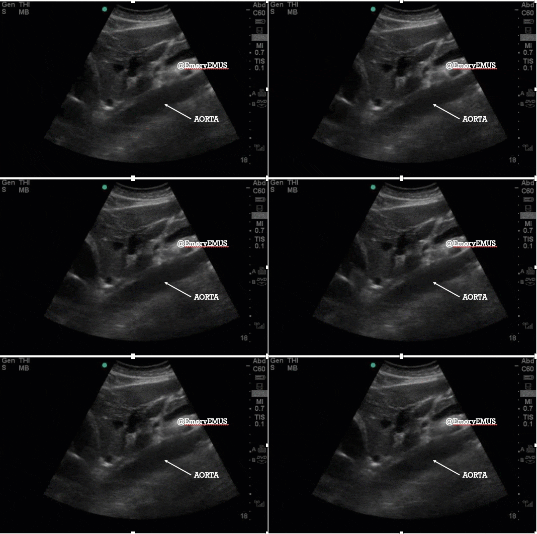
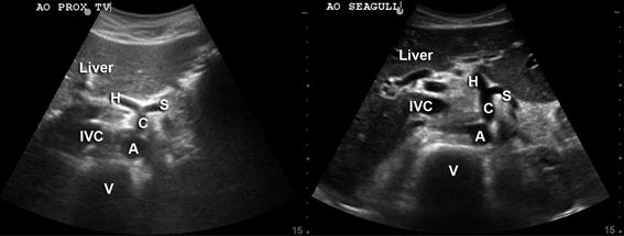
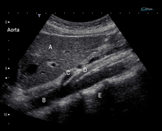
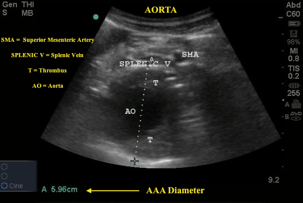
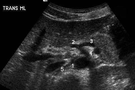
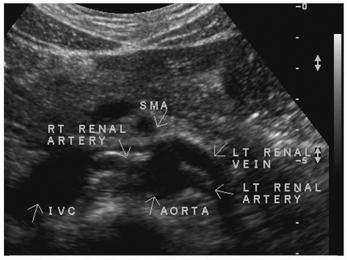
Aorta branches ultrasound. An abdominal ultrasound is done to view structures inside the abdomen. Our goal in aortic ultrasound is to visualize the entirity of the abdominal aorta beginning at the level of the celiac artery all the way to its bifurcation into the common iliac arteries. The major branches of the abdominal aorta include the celiac axis the superior mesenteric artery the renal arteries the inferior mesenteric artery the gonadal arteries and the lumbar arteries. An abdominal aortic aneurysm occurs when a lower portion of your bodys main artery aorta becomes weakened and bulges.
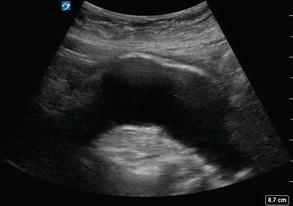
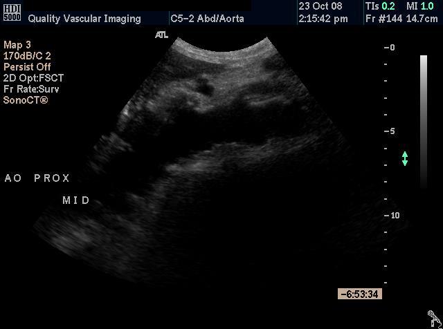
The abdominal aorta is the major conduit artery distributing blood to the abdominal organs and then to the lower extremities. It is divided into. Notice as you scan distally the aorta becomes more superficial or anterior in its location and its diameter tapers before the bifurcation. Scan the length of the aorta from proximal to distal until you reach the aortoiliac bifurcation.
We will measure the diameter of the aorta at 3 different levels to assess for any areas of dilatation.





1.jpg)



















.jpg)


